ROBOTIC SURGICAL SYSTEM
Today, thanks to the da Vinci® Robotic Surgery System, surgeons can perform important surgeries by intervening on the patient through small holes. This opportunity, gained by humanity with the development of medical technologies, provides great comfort and convenience to the surgeon and the patient. In our hospital, ENT, General Surgery, Gynecology and Urological Radical Cancer Surgery are performed with Da Vinci Robotic Surgery.
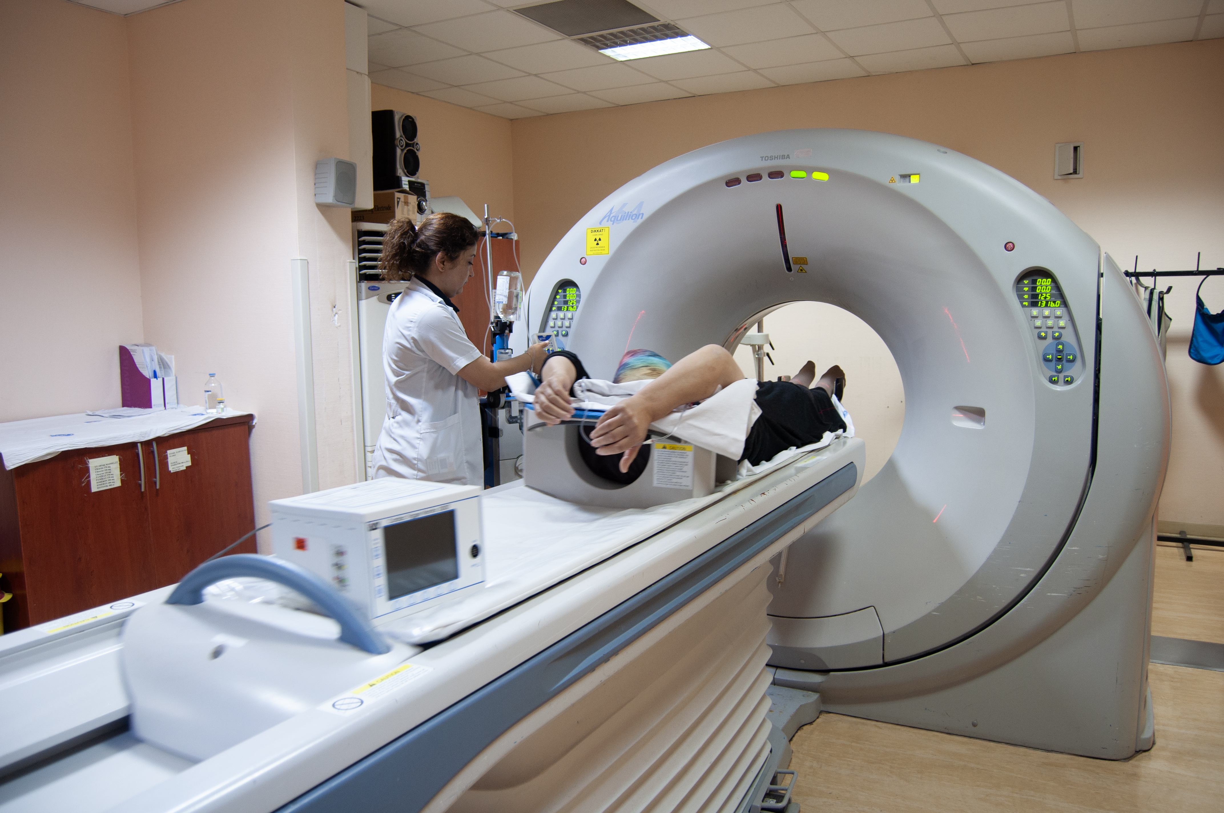
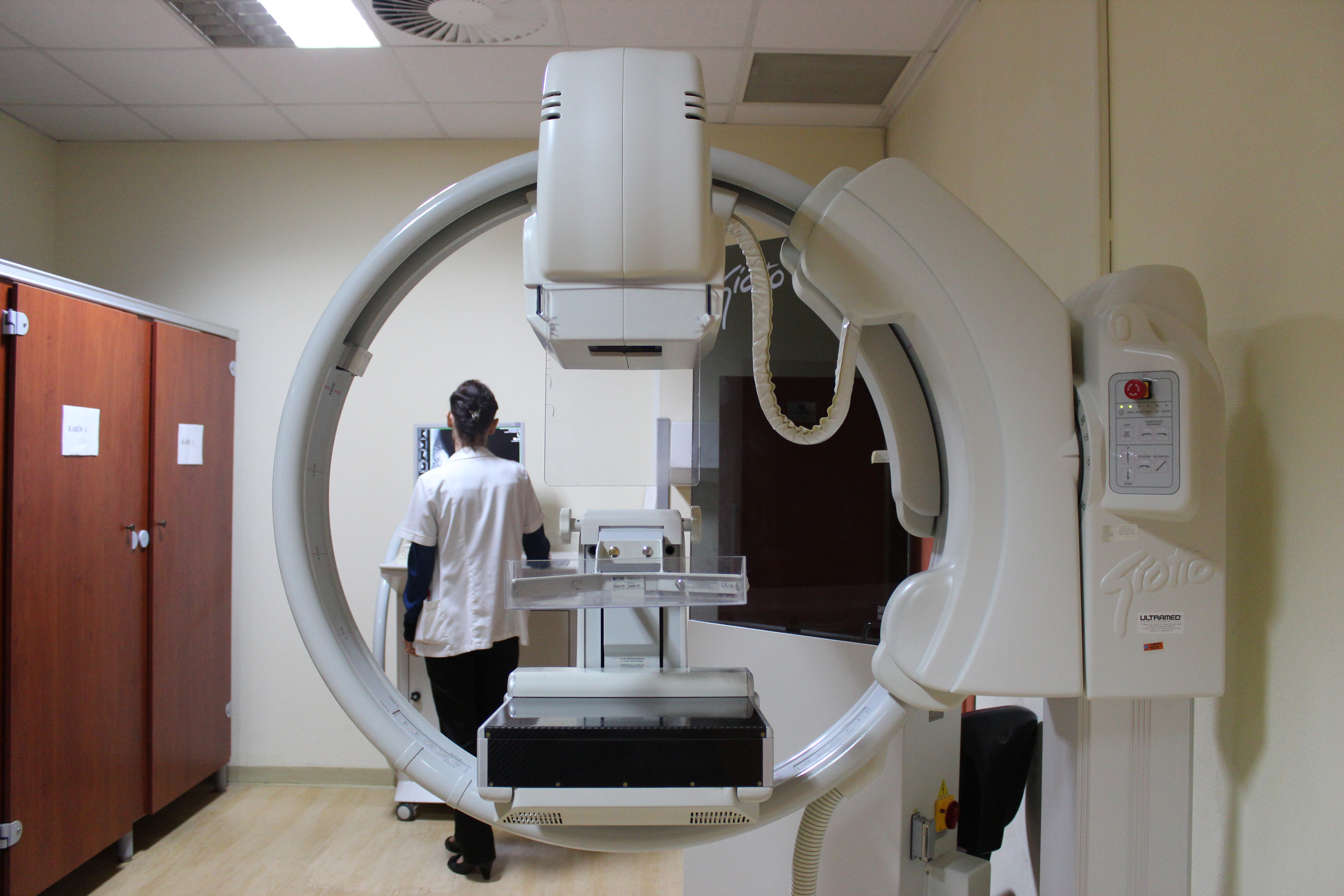
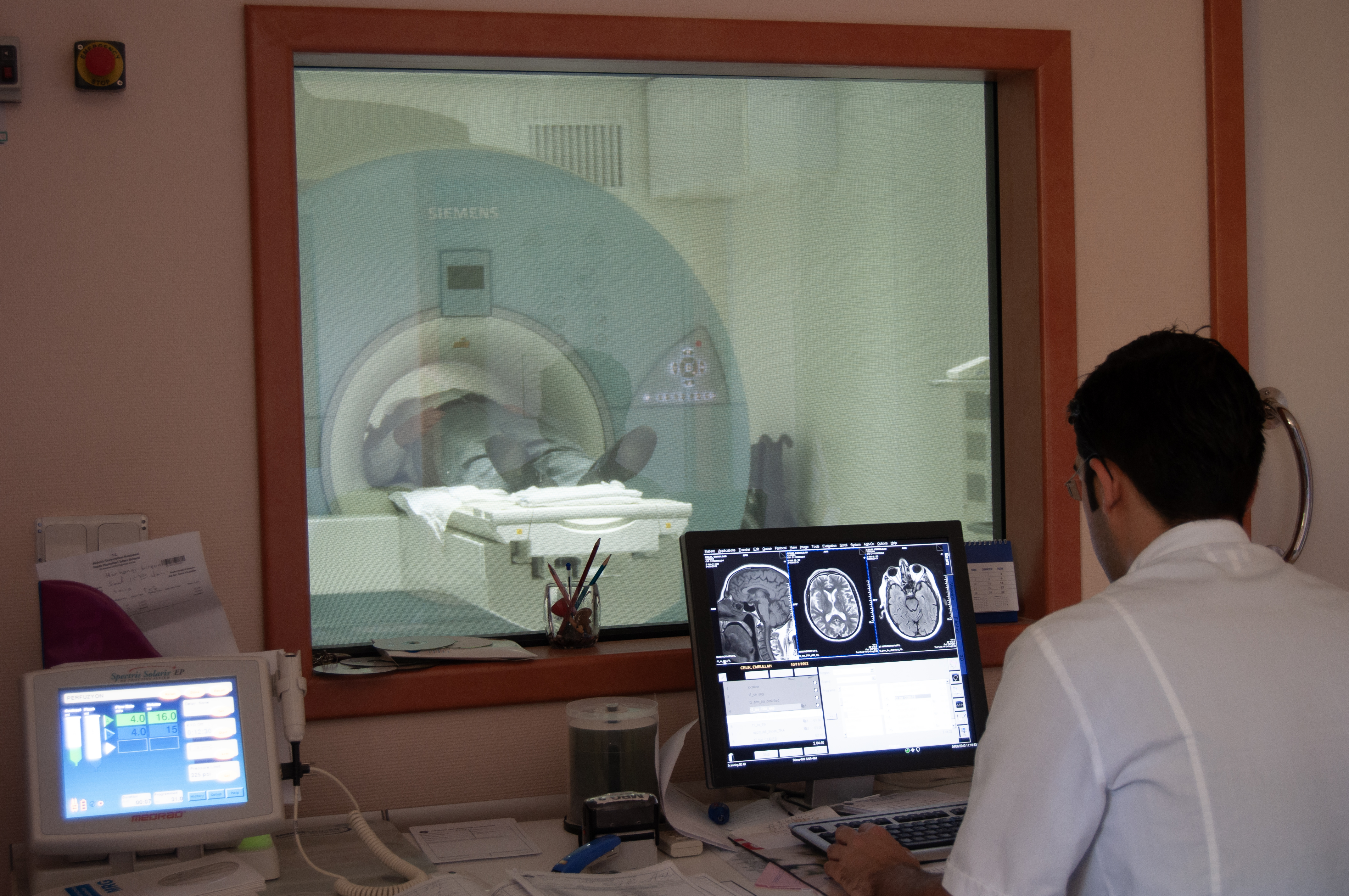
World-class diagnostic examinations and imaging-guided biopsy procedures are performed in our radiology department. The department equipped with four magnetic resonance imaging (MRI) units, three computed tomography (CT) units, two angiography units, two digital mammography units, 12 Doppler ultrasonography devices, as well as a fluoroscopy unit and many digital x-ray machines.
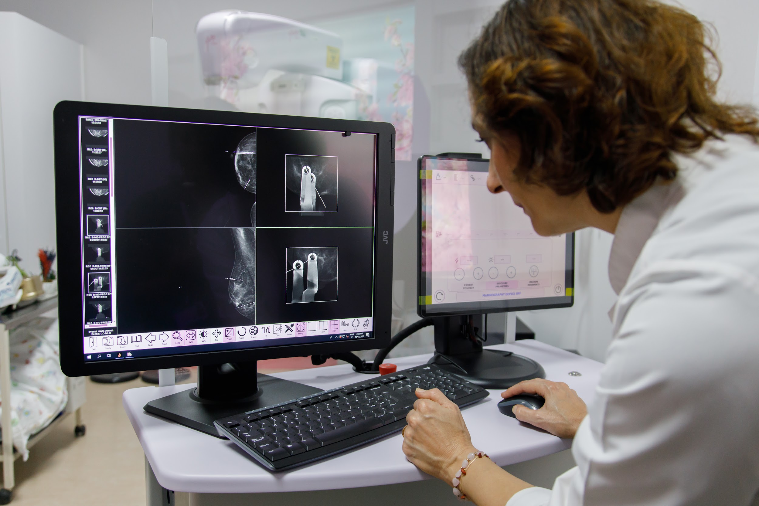
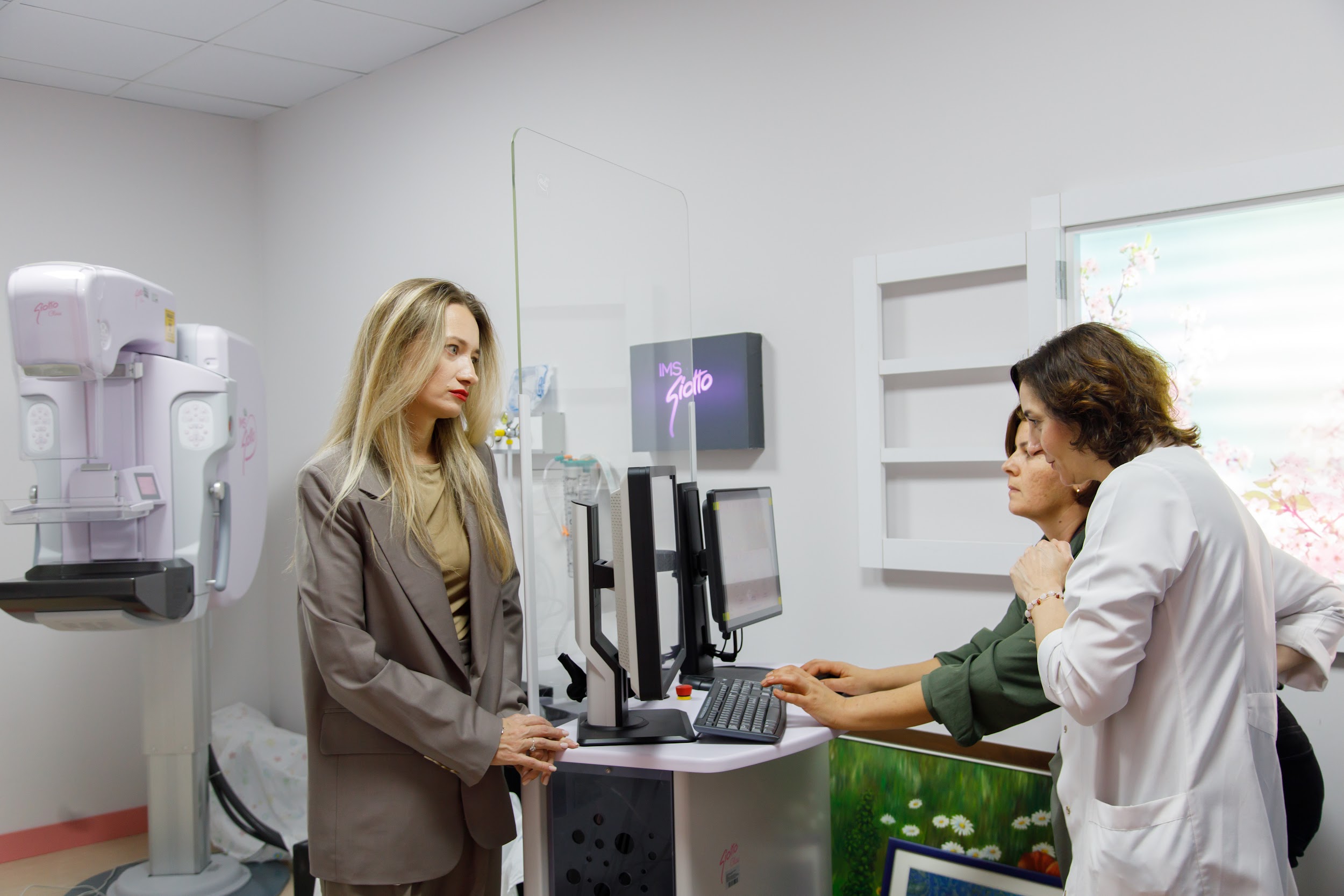
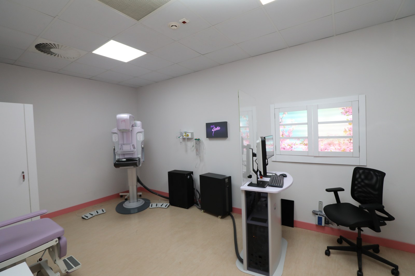
Breast Health and Diseases Center was opened at Akdeniz University Hospital, a first in public institutions in Turkey.Diagnoses applied in our center;digital mammography,digital tomosynthesis of the breast,Contrast-enhanced mammography,Ultrasound examination of the mammary gland,magnetic resonance imaging (MRI) of the mammary gland.Associate Professor Ebru Sanhal carries out the following studies professionally:Vacuum biopsy under ultrasound/tomosynthesis/contrast mammography control,core needle biopsy (tru-cut biopsy / cortical biopsy),Wire localization under ultrasound/mammography control, Marking the tumor and axillary lymph nodes with clips/markers before chemotherapy in patients diagnosed with breast cancer, Interventional procedures for diagnostic and therapeutic purposes under imaging control(fine needle aspiration biopsy, abscess drainage).- Interventional procedures for diagnostic and therapeutic purposes under imaging control (fine needle aspiration biopsy, abscess drainage).
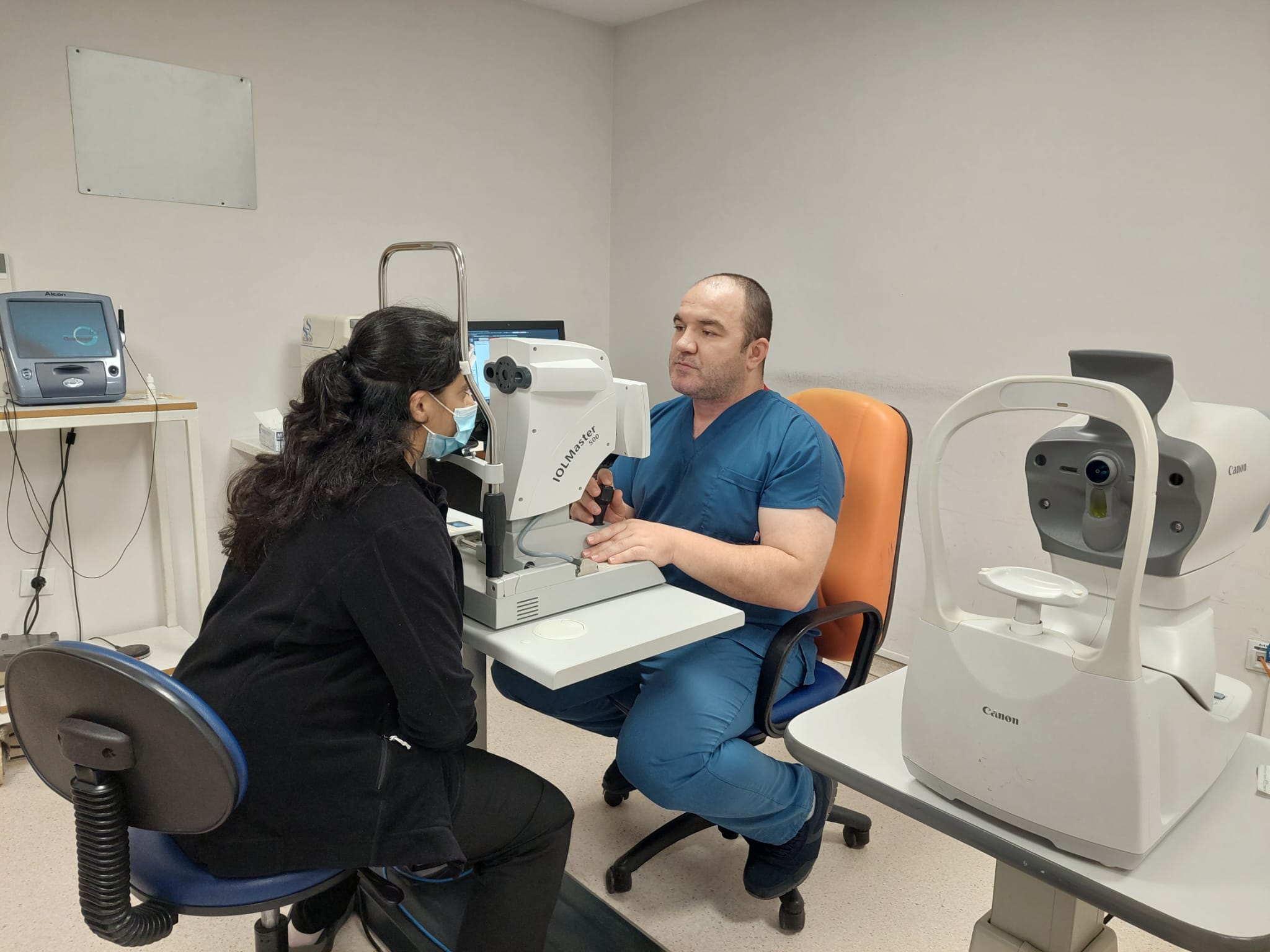
Iol Master 500: Millions of people around the world undergo cataract surgery every year. Calculation of the intraocular lens (IOL) before the eye surgery is done with IOL Master (Optical Biometry) or A-Scan (Ultrasonic Biometry) devices. IOLmaster 500 is a very important device that enables the calculation of the power of the intraocular lens (IOL) placed in the eye during cataract surgery, without optically touching the eye.
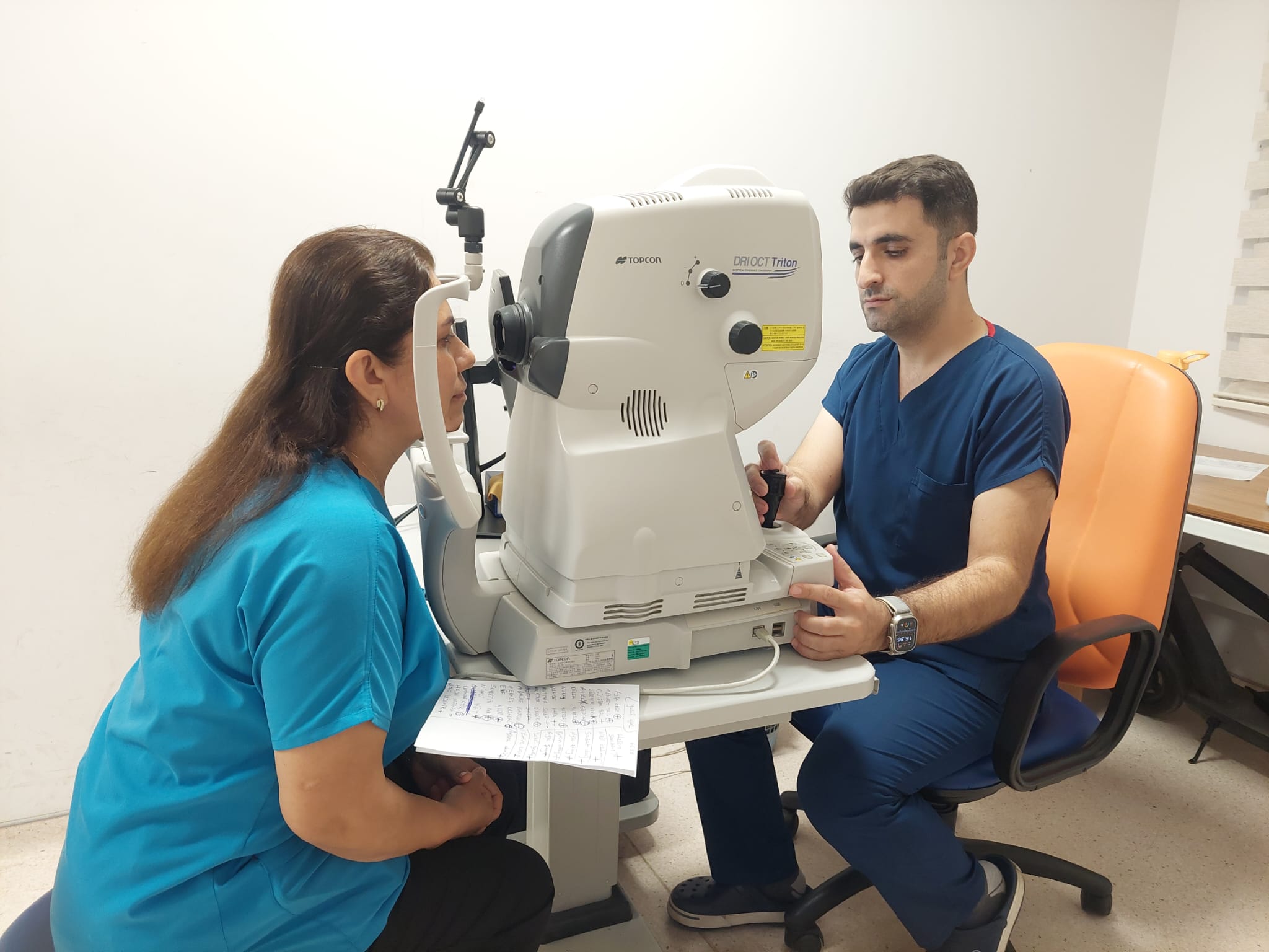
Topcon Dri Oct Triton : Non-invasive diagnostic method that enables examination by taking high-resolution sections of tissues. OCT which can generally be used for abnormalities of both the posterior segment of the eye and the anterior segment, provides a modern contribution to the diagnosis and treatment of many diseases in our clinic. We can analyze retina and macular thickness, vitreomacular shrinkages, pre-retinal membranes (epiretinal membranes), macular holes, macular edema, damage caused by diabetes and vascular occlusions, and pathologies in the cornea.
Ocular Ultrasonography: Ocular (eye) ultrasonography; It is a diagnostic method that allows imaging of the intraocular lens, vitreous, retina and soft tissues around the eye. It is an easy-to-perform, painless, non-invasive examination that provides information about the posterior segment of the eye and especially the retina, using the retroreflective properties of tissues with the help of sound waves.It is useful in the evaluation of intraocular structures in cases of environmental turbidity such as cataract and intraocular bleeding, where intraocular structures cannot be evaluated by examination, and in the detection and follow-up of retinal detachment, intraocular foreign bodies and intraocular tumors.
Anterior Segment Photography: It enables the detection of pathologies related to the structures in the front part of the eye such as sclera, conjunctiva, cornea, iris and lens, and objective visualization of the changes over time. It is especially useful in detecting pre- and postoperative changes.
.jpeg)
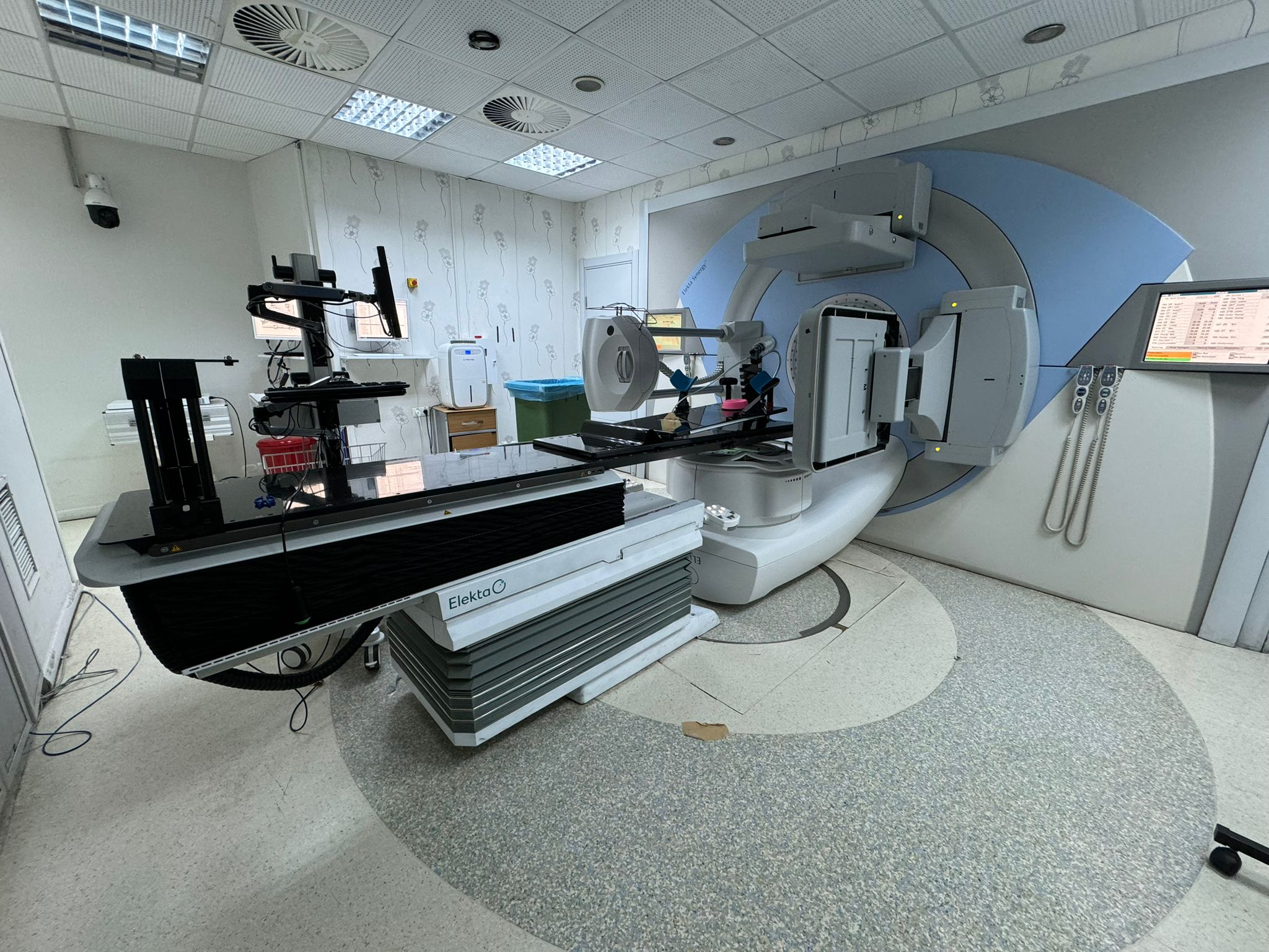
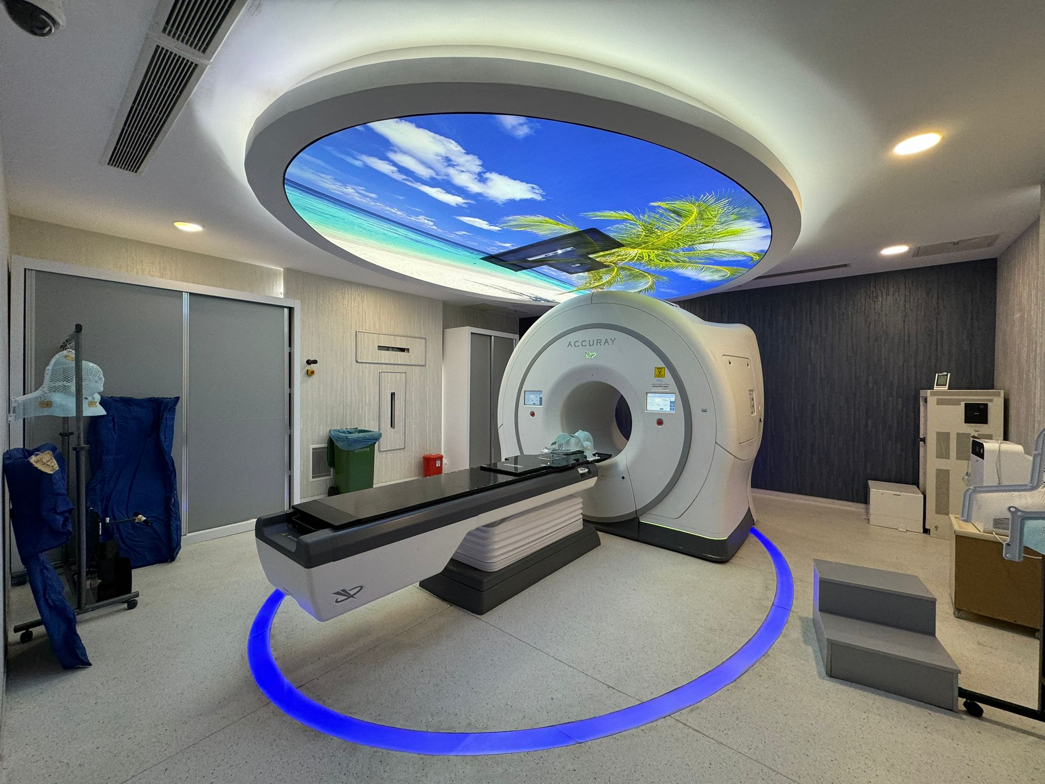
Akdeniz University is one of our country's leading universities that has pioneered vital awareness in organ transplantation and has become one of the world's distinguished organ transplantation centers through its educational and research services in this field, conducting multi-organ and composite tissue transplants within its facilities.
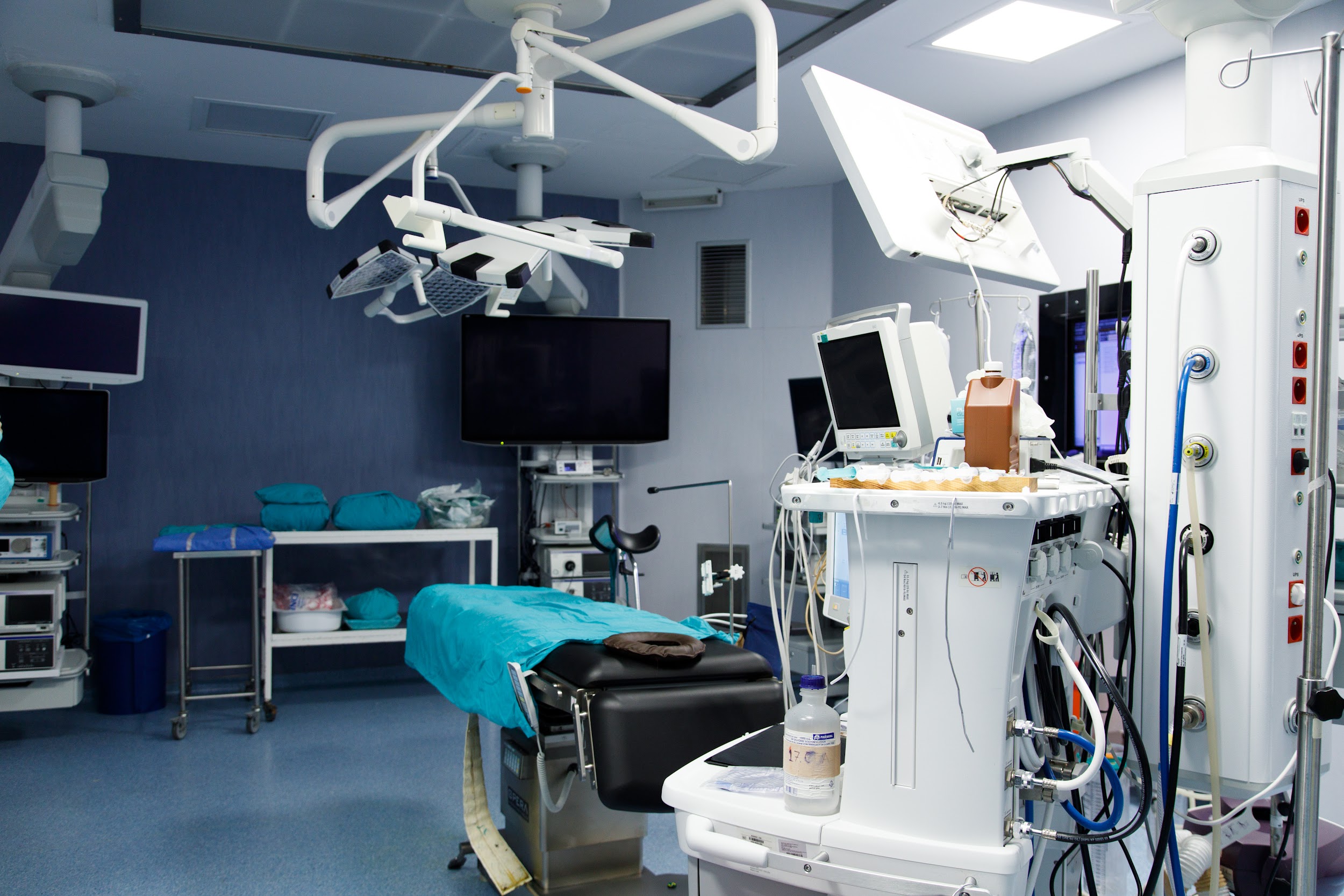
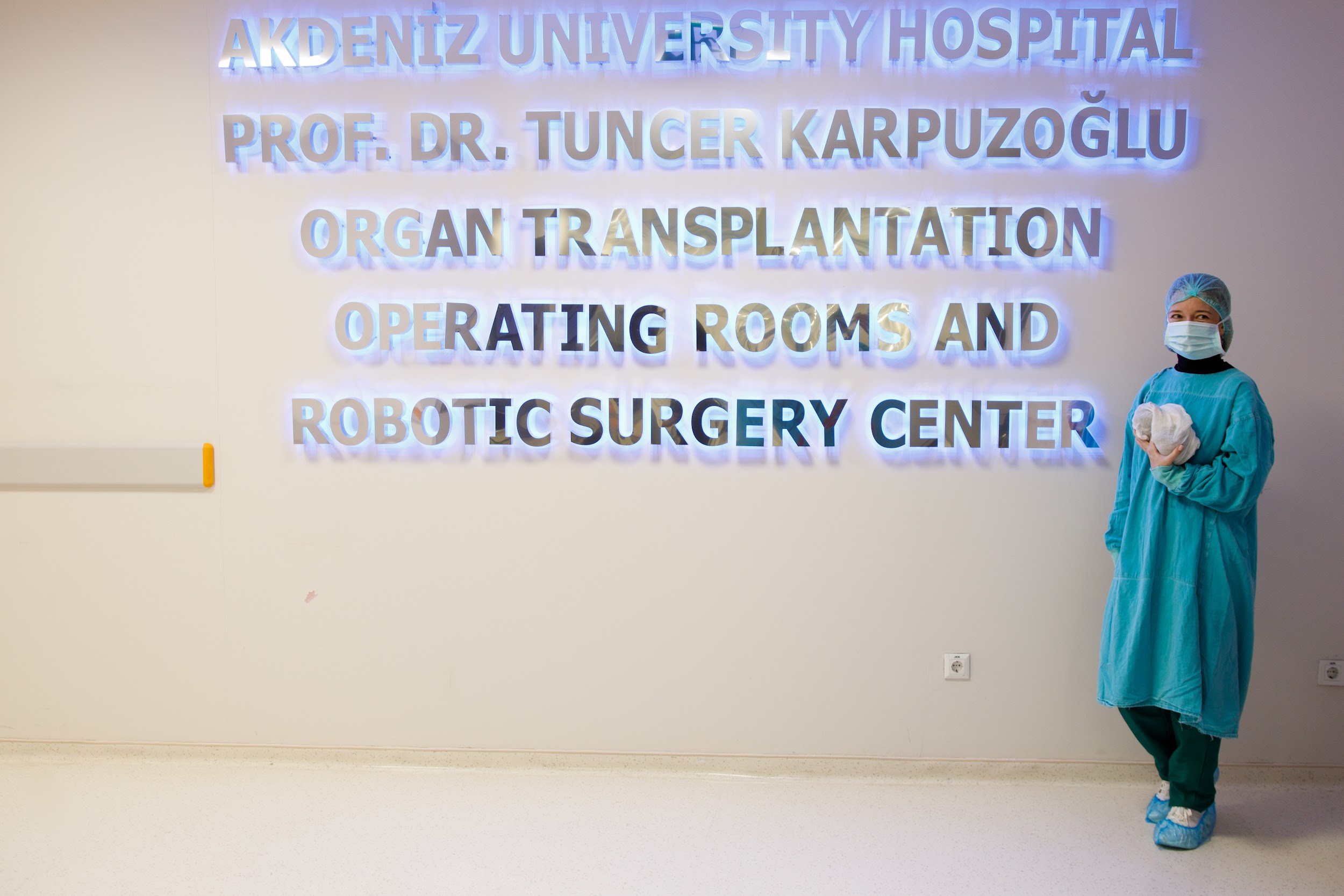
There are 3 coronary angiography devices in the cardiology department. In our angiography unit, percutaneous coronary intervention, electrophysiological study, pacemaker implantation, pericardiocentesis, valve interventions and peripheral vascular interventions are performed. In addition, non-invasive evaluations are performed in our department for the diagnosis of cardiovascular diseases using echocardiography, cardiovascular stress test (exertion test), TILT test (tilt table test), rhythm and blood pressure holter monitoring devices.
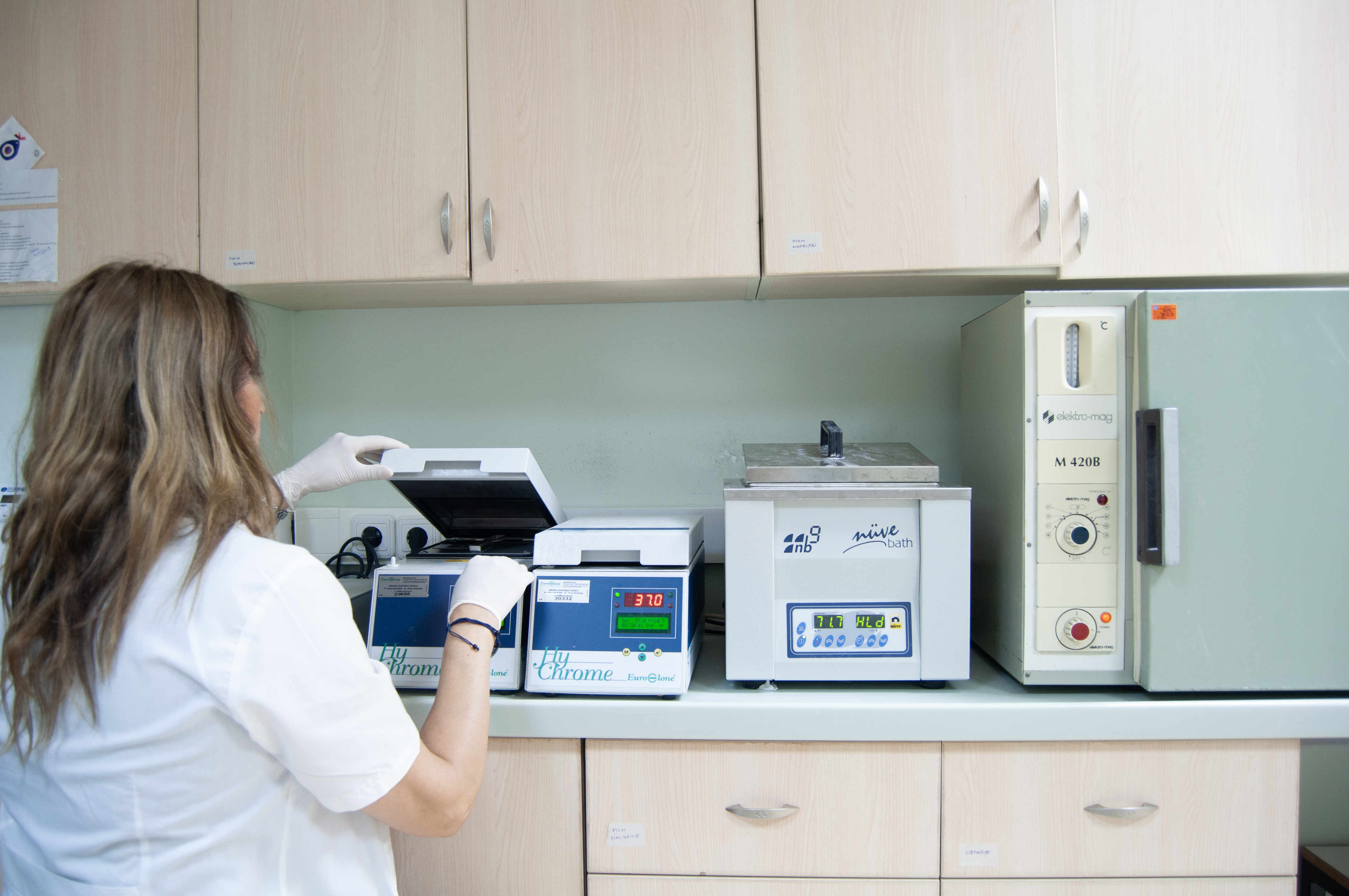
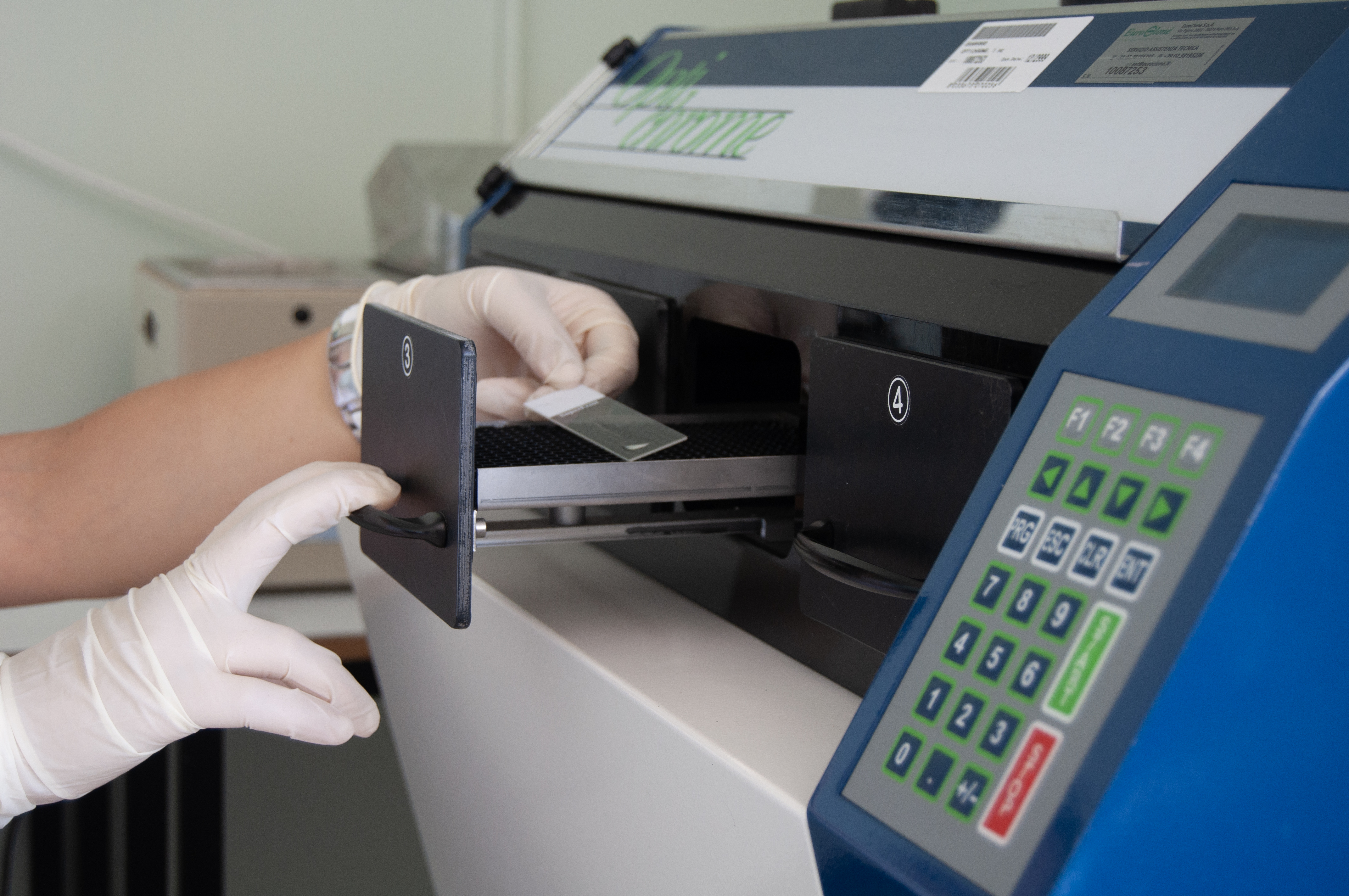
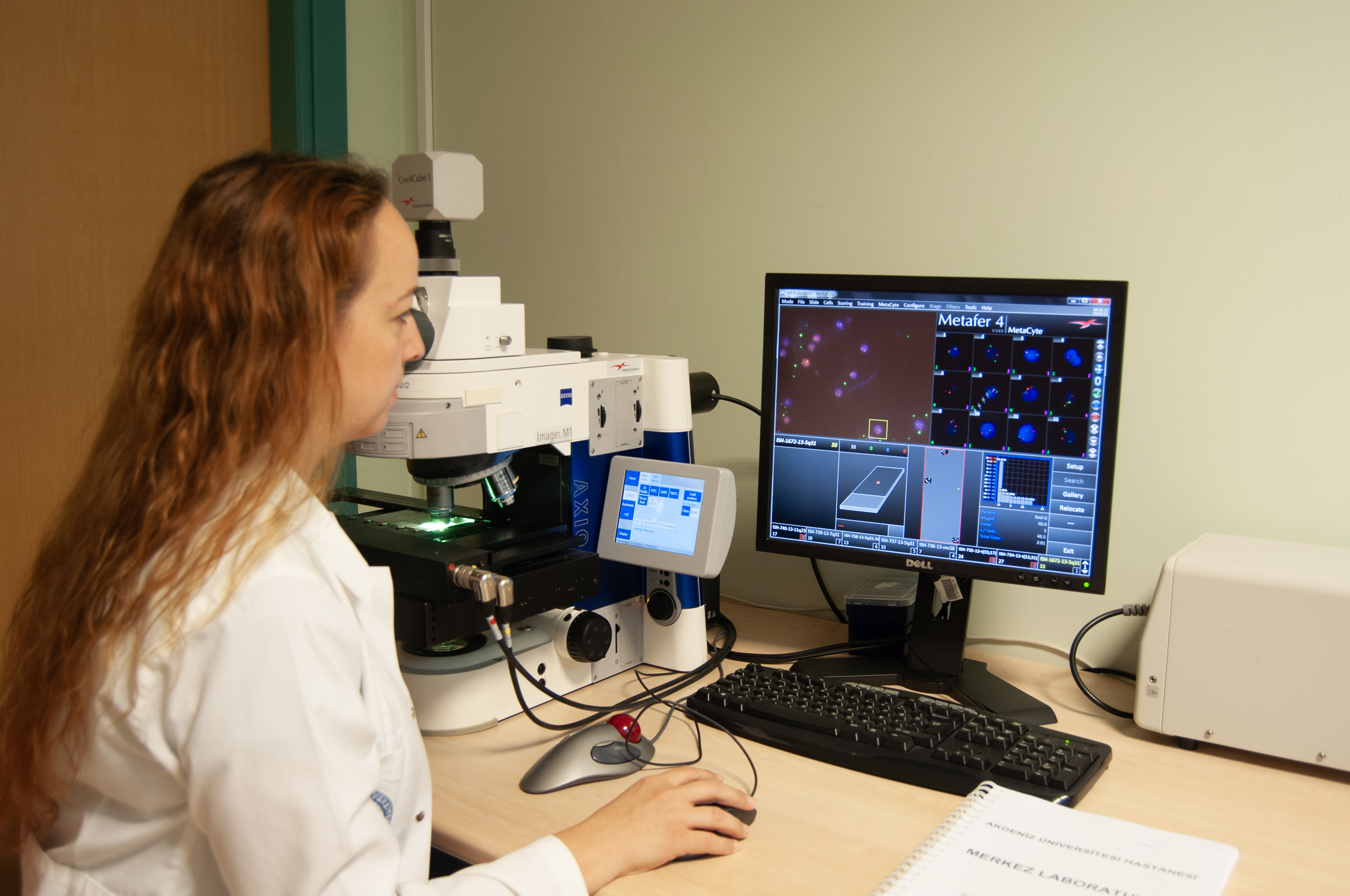
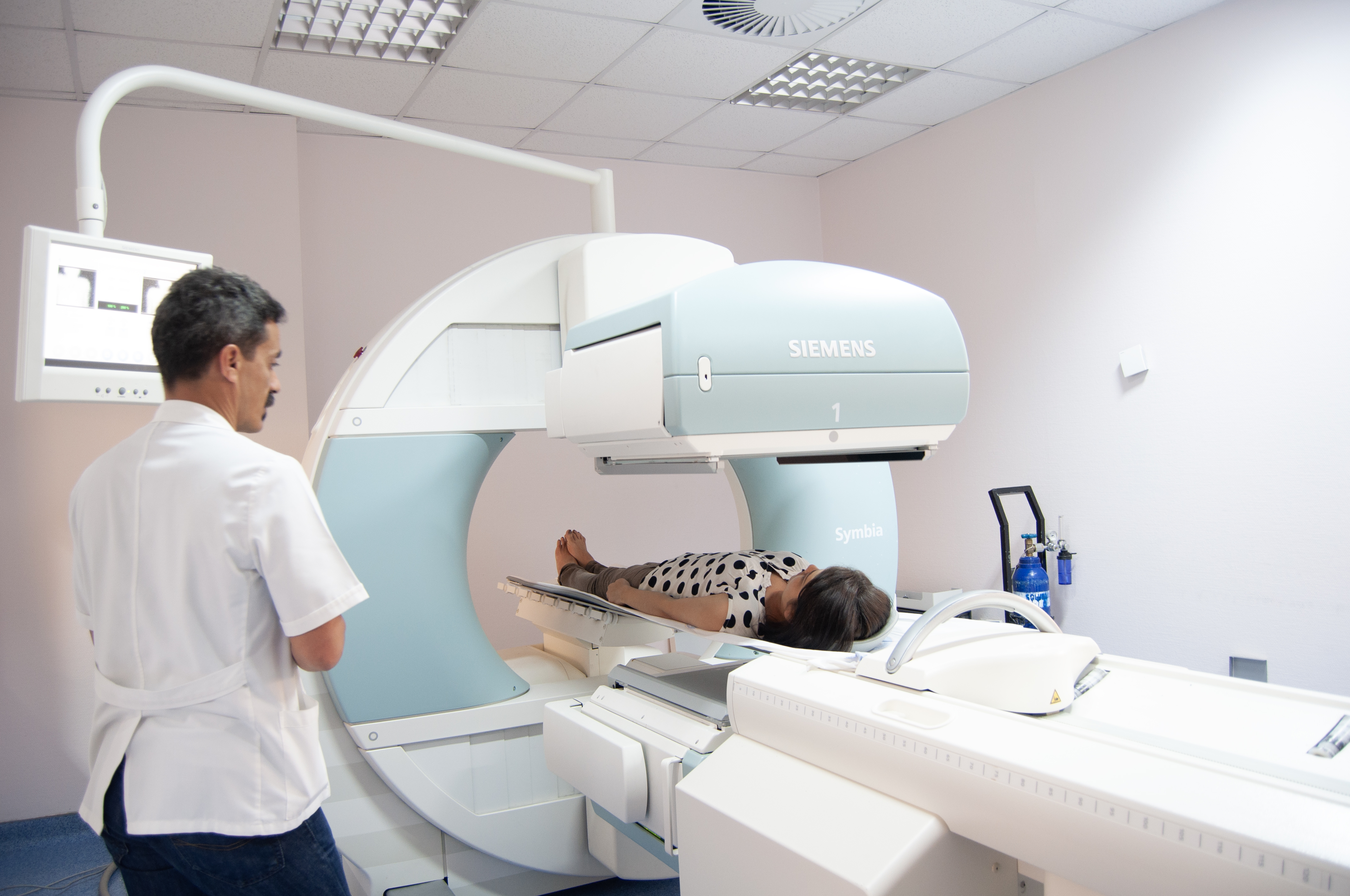
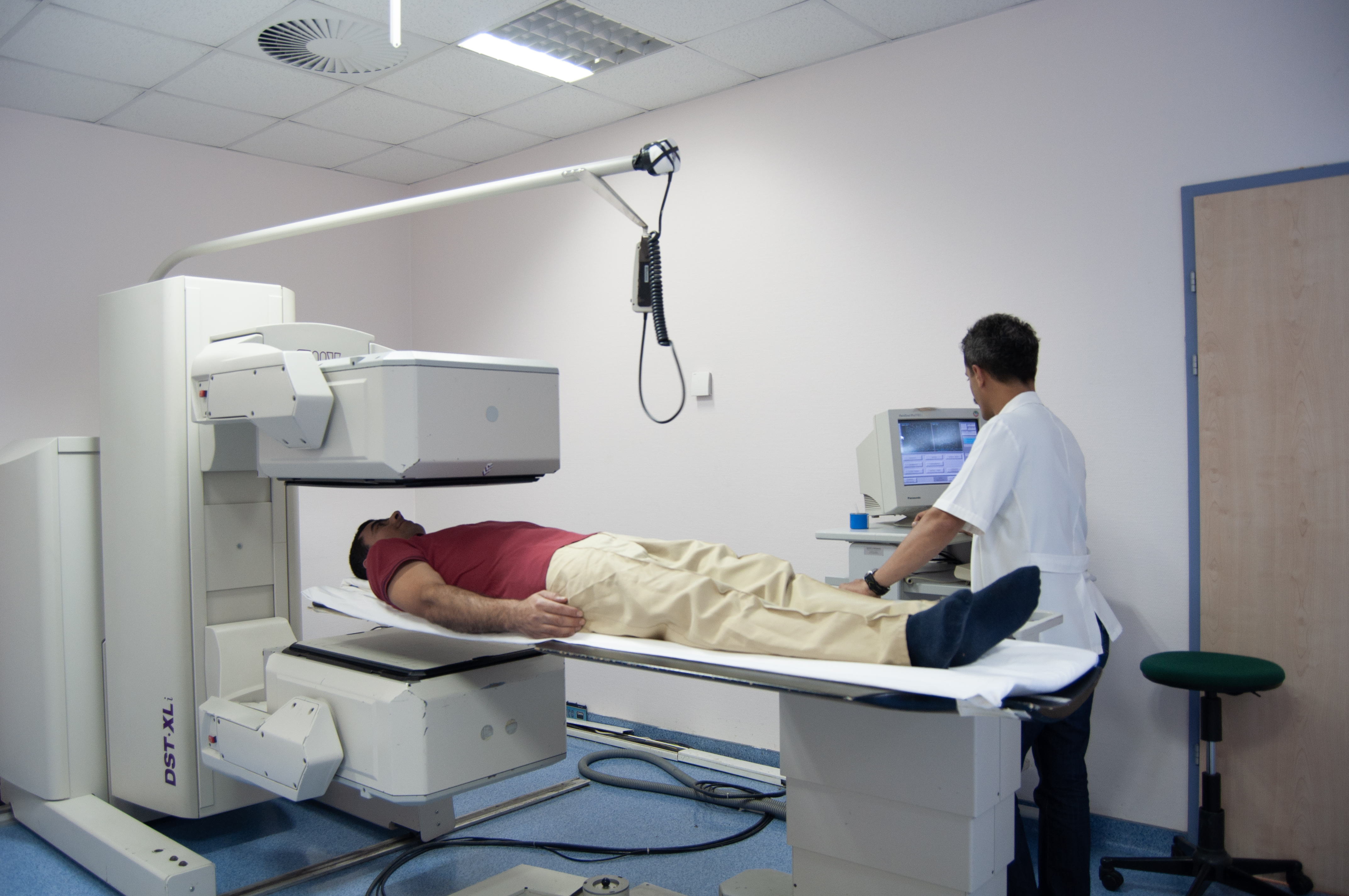
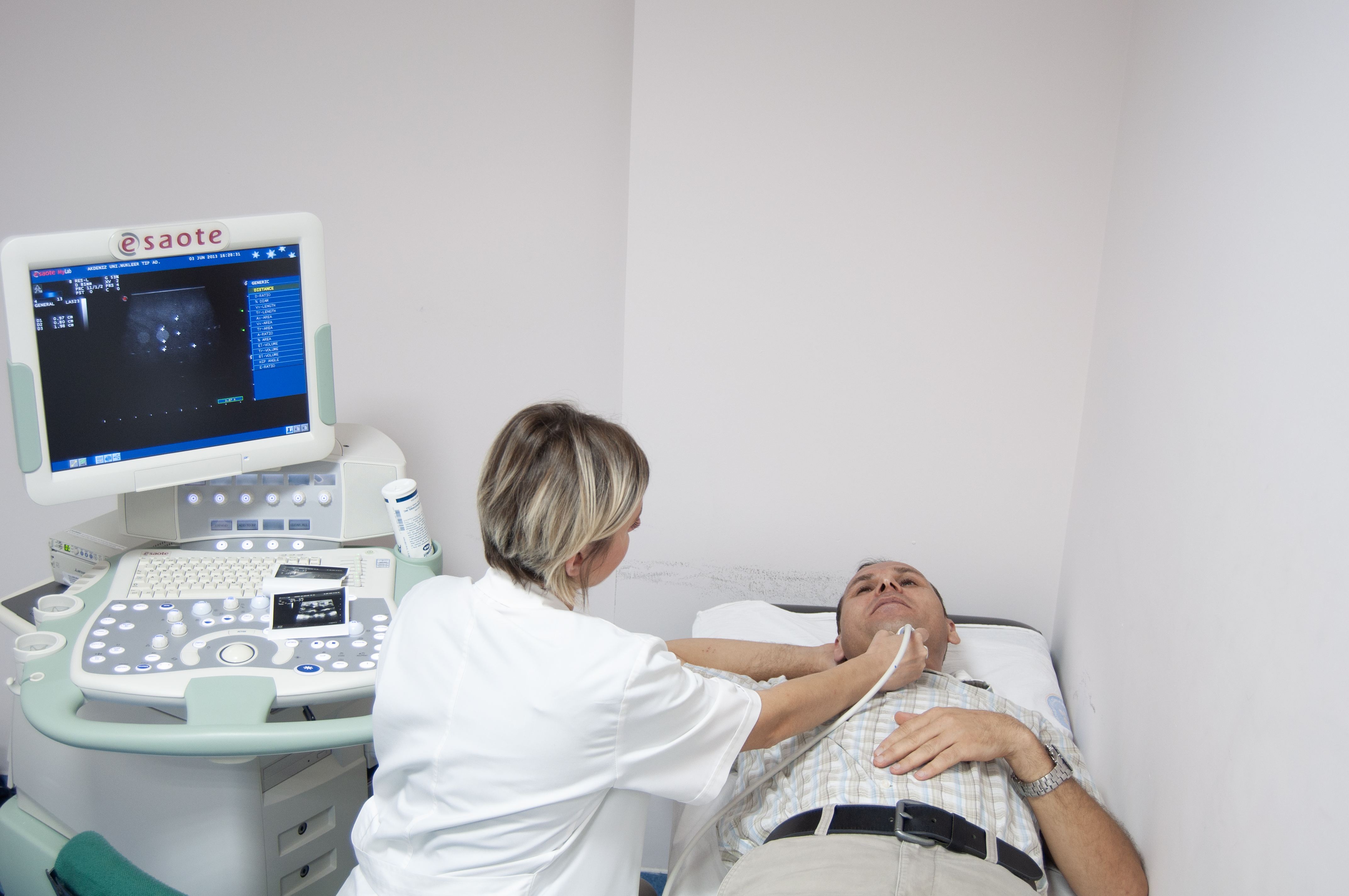
Our center is a referral hospital providing comprehensive clinical services in radioisotope imaging and therapy, serving the Mediterranean region and other neighboring countries including the European Union, the Russian Federation, and Middle Eastern countries. Equipped with True D technology, we have a Positron Emission Tomography (PET)/CT scanner, two Single Photon Emission Computed Tomography (SPECT) scanners, and Dual-energy X-ray Absorptiometry (DEXA) for Bone Mineral Density Measurement, Ultrasound for Ultrasonography.
Son güncelleme : 23.08.2024 16:41:20
